Preprint. A sensitive orange fluorescent calcium ion indicator for imaging neural activity
A. Aggarwal, H.A. Baker, C.D. Dürst, I-W. Chen, P. de Chambrier, J.M. Gonzales, J.S. Marvin, M. Vandal, T. Lundberg, K. Sakoi, R. Patel, C-Y. Wang, F. Visser, Y. Fouad, S. Sunil, M. Wiens, T. Terai, K. Takahashi-Yamashiro, R.J. Thompson, T.A. Brown, Y. Nasu, M.D. Nguyen, G.R.J. Gordon, S. McFarlane, K. Podgorski, A. Holtmaat, R.E. Campbell and A.W. Lohman*, “A sensitive orange fluorescent calcium ion indicator for imaging neural activity”, submitted. Preprint posted to bioRxiv 2025.07.28.667269.
Preprint. High-performance genetically-encoded green and red fluorescent biosensors for pyruvate
S. Imai (equal contribution), S. Hario (equal contribution), C. Suerte, I. Yamaguchi, K. Sakoi, T. Terai, K. Takahashi-Yamashiro, and R.E. Campbell*, “High-performance genetically-encoded green and red fluorescent biosensors for pyruvate”. Preprint posted to bioRxiv 2025.04.17.649293.
Preprint. PinkyCaMP: a mScarlet-based calcium sensor with exceptional brightness, photostability, and multiplexing capabilities
R. Fink (equal contribution), S. Imai (equal contribution), N. Gockel, G. Lauer, K. Renken, J. Wietek, P.J. Lamothe-Molina, F. Furhmann, M. Mittag, T. Ziebarth, A. Canziani, M. Kubitschke, V. Kistmacher, A. Kretschmer, E. Sebastian, D. Schmitz, T. Terai, J. Gruendemann, S. Hassan, T. Patriarchi, A. Reiner, M. Fuhrmann, R.E. Campbell, and O.A. Masseck*, “PinkyCaMP: a mScarlet-based calcium sensor with exceptional brightness, photostability, and multiplexing capabilities”, submitted. Preprint posted to bioRxiv 2024.12.16.628673.
157. A red fluorescent genetically encoded biosensor for in vivo imaging of extracellular L-lactate dynamics
Y. Kamijo, P. Mächler (equal contribution), N. Ness (equal contribution), C.Q. Vu (equal contribution), T. Kusakizako (equal contribution), J. Mannuthodikayil, Z. Ku, M. Boisvert, E. Grebenik, I. Miyazaki, R. Hashizume, H. Sato, R. Liu, Y. Hori, T. Tomita, T. Katayama, A. Furube, G. Caraveo, M-E. Paquet, M. Drobizhev, O.Nureki, S.Arai, M. Brancaccio, R.E. Campbell, D. Kleinfeld, and Y. Nasu*, “A red fluorescent genetically encoded biosensor for in vivo imaging of extracellular L-lactate dynamics”, Nat. Commun., 2025, 16, 9531. Preprint posted to bioRxiv 2022.08.30.505811.
156. Bipartite Genetically Encoded Biosensors to Sense Calcium Ion Dynamics at Membrane-Membrane Contact Sites
I. Yamaguchi (equal contribution), L. Barazzuol (equal contribution), G. Dematteis, W. Zhu, Y. Wen, M. Drobizhev, D. Lim, R.E. Campbell, T. Calì* and Y. Nasu*, “Bipartite Genetically Encoded Biosensors to Sense Calcium Ion Dynamics at Membrane-Membrane Contact Sites”, Anal. Chem., 2025, 97, 19848–19861.

155. Far-red fluorescent genetically encoded calcium ion indicators
R. Dalangin, B.Z. Jia, Y. Qi, A. Aggarwal, K. Sakoi, M. Drobizhev, R.S. Molina, R. Patel, A.S. Abdelfattah, J. Zheng, D. Reep, J.P. Hasseman, The GENIE Project Team, Y. Zhao, J. Wu, K. Podgorski, A.G. Tebo, E.R. Schreiter, T.E. Hughes, T. Terai, M-E. Paquet, S.G. Megason, A.E. Cohen, Y. Shen*, and R.E. Campbell*, “Far-red fluorescent genetically encoded calcium ion indicators”, Nat. Commun., 2025, 16, 3318. Preprint posted to bioRxiv 2020.11.12.380089.
154. Synthesis and application of a photocaged-L-lactate for studying the biological roles of L-lactate
I. Miyazaki, K.K. Tsao*, Y. Kamijo, Y. Nasu, T. Terai*, R.E. Campbell*, “Synthesis and application of a photocaged-L-lactate for studying the biological roles of L-lactate”, Commun. Chem., 2025, 8, 104. Preprint posted to bioRxiv 2024.01.30.577898.

153. Astrocyte Kir4.1 expression level territorially controls excitatory transmission in the brain
O. Tyurikova*, O. Kopach, K. Zheng, D. Rathore, N. Codadu, S-Y. Wu, Y. Shen, R.E. Campbell, R.C. Wykes, K. Volynski, L.P. Savtchenko, and D.A. Rusakov*, “Astrocyte Kir4.1 expression level territorially controls excitatory transmission in the brain”, Cell Rep., 2025, 44, 115299.
152. A chemigenetic indicator based on a synthetic chelator and a green fluorescent protein for imaging of intracellular sodium ion
S. Takeuchi, S. Imai, T. Terai*, and R.E. Campbell*, “A chemigenetic indicator based on a synthetic chelator and a green fluorescent protein for imaging of intracellular sodium ion”, RSC Chem. Biol., 2025, 6, 170-174.

151. Multimodal fluorescence-optoacoustic in vivo imaging of the near-infrared calcium ion indicator NIR-GECO2G
S.F Shaykevich, J.P. Little, Y. Qian, M-E. Paquet, R.E. Campbell*, D. Razansky*, and S. Shoham*, “Multimodal fluorescence-optoacoustic in vivo imaging of the near-infrared calcium ion indicator NIR-GECO2G”, Photoacoustics, 2025, 41, 100671.
150. A toolbox for ablating excitatory and inhibitory synapses
A. Bareghamyan, C. Deng, S. Daoudi, S.C. Yadav, X. Lu, W. Zhang, R.E. Campbell, R.H. Kramer, D.M. Chenoweth, and D.B Arnold*, “A toolbox for ablating excitatory and inhibitory synapses”, eLife, 2024, 13, RP103757. Preprint posted to bioRxiv 2024.09.23.614589.
149. High-performance chemigenetic potassium ion indicator
D. Cheng, Z. Ouyang, X. He, Y. Nasu, Y. Wen, T. Terai*, and R.E. Campbell*, “High-performance chemigenetic potassium ion indicator”, J. Am. Chem. Soc., 2024, 146, 35117-35128.

148. High-Throughput Discovery of Substrate Peptide Sequences for E3 Ubiquitin Ligases Using a cDNA Display Method
K. Tamagawa, R.E. Campbell, and T. Terai*, “High-Throughput Discovery of Substrate Peptide Sequences for E3 Ubiquitin Ligases Using a cDNA Display Method”, ChemBioChem, 2024, 25, e202400617.

147. The best of both worlds: Chemigenetic fluorescent sensors for biological imaging
K.K. Tsao* (equal contribution), S. Imai (equal contribution), M. Chang, S. Hario, T. Terai*, and R.E. Campbell*, “The best of both worlds: Chemigenetic fluorescent sensors for biological imaging”, Cell Chem. Biol., 2024, 31, 1652-1664.

146. A lactate-dependent shift of glycolysis mediates synaptic and cognitive processes in male mice
I. Fernández-Moncada*, G. Lavanco, U.B. Fundazuri, N. Bollmohr, S. Mountadem, T. Dalla Tor, P. Hachaguer, F. Julio-Kalajzic, D. Gisquet, R. Serrat, L. Bellocchio, A. Cannich, B. Fortunato-Marsol, Y. Nasu, R.E. Campbell, F. Drago, C. Cannizzaro, G. Ferreira, A-K. Bouzier-Sore, L. Pellerin, J.P. Bolaños, G. Bonvento, L.F. Barros, S.H.R. Oliet, A. Panatier, and G. Marsicano*, “A lactate-dependent shift of glycolysis mediates synaptic and cognitive processes in male mice”, Nat. Commun., 2024, 15, 6842. Preprint posted to bioRxiv 2023.03.15.532748.
145. An automated screening platform for improving the performance of genetically encoded Ca2+ biosensors in mammalian cells
Y. Zhao*, Y. Shen, T. Veres, and R.E. Campbell*, “An automated screening platform for improving the performance of genetically encoded Ca2+ biosensors in mammalian cells”, Sens. Diagn., 2024, 3, 1494-1504.
144. Development of an miRFP680-Based Fluorescent Calcium Ion Biosensor Using End-Optimized Transposons
F. Chai, H. Fujii (equal contribution), G.N.T. Le (equal contribution), C. Lin (equal contribution), K. Ota (equal contribution), K.M. Lin, L.M.T. Pham, P. Zou, M. Drobizhev, Y. Nasu, T. Terai, H. Bito, and R.E. Campbell*, “Development of an miRFP680-Based Fluorescent Calcium Ion Biosensor Using End-Optimized Transposons”, ACS Sens., 2024, 9, 3394–3402.

143. An optogenetic method for the controlled release of single molecules
P. Kashyap, S. Bertelli, F. Cao, Y. Kostritskaia, F. Blank, N.A. Srikanth, C. Schlack-Leigers, R. Saleppico, D. Bierhuizen, X. Lu, W. Nickel, R.E. Campbell, A.J.R. Plested, T. Stauber, M.J. Taylor, and H. Ewers*, “An optogenetic method for the controlled release of single molecules”, Nat. Methods, 2024, 21, 666–672. Preprint posted to bioRxiv 2023.09.16.557871.
142. A blue-shifted genetically encoded Ca2+ indicator with enhanced two-photon absorption
A. Aggarwal, S. Sunil (equal contribution), I. Bendifallah (equal contribution), M. Moon, M. Drobizhev, L. Zarowny, J. Zheng, S.-Y. Wu, A.W. Lohman, A.G. Tebo, V. Emiliani, K. Podgorski, Y. Shen*, and R.E. Campbell*, “A blue-shifted genetically encoded Ca2+ indicator with enhanced two-photon absorption”, Neurophoton., 2024, 11, 024207. Preprint posted to bioRxiv 2023.10.12.562058.
141. High performance genetically-encoded green fluorescent biosensors for intracellular L-lactate
S. Hario (equal contribution), G.N.T. Le (equal contribution), H. Sugimoto, K. Takahashi-Yamashiro, S. Nishinami, H. Toda, S. Li, J.S. Marvin, S. Kuroda, M. Drobizhev, T. Terai, Y. Nasu*, and R.E. Campbell*, “High performance genetically-encoded green fluorescent biosensors for intracellular L-lactate”, ACS Cent. Sci., 2024, 10, 402–416. Preprint posted to bioRxiv 2022.10.19.512892.

140. 蛍光タンパク質とタグタンパク質 [Fluorescent proteins and protein tags]
J. Adachi, T. Terai, S. Hario, R.E. Campbell, and Y. Hori, “蛍光タンパク質とタグタンパク質 (Fluorescent proteins and protein tags)”, 現代化学 (Chemistry Today), 2024, 640, 33-39. (Not peer-reviewed)
139. タンパク質を基盤とする蛍光イメージングセンサー開発の最前線 [Development of fluorescent imaging sensors based on proteins]
針尾 紗彩, 竹内 志織, キャンベル ロバート アール*, 寺井 琢也*, “タンパク質を基盤とする蛍光イメージングセンサー開発の最前線“, 日本薬理学雑誌, 2024, 159, 25-30. [Original citation]
S. Hario, S. Takeuchi, R.E. Campbell*, and T. Terai*, “Development of fluorescent imaging sensors based on proteins”, Folia Pharmacol. Jpn., 2024, 159, 25-30. [Translated citation]
138. A monochromatically excitable green-red dual-fluorophore fusion incorporating a new large Stokes shift fluorescent protein
J.O. Ejike (equal contribution), M. Sadoine (equal contribution), Y. Shen (equal contribution), Y. Ishikawa (equal contribution), E. Sunal, S. Hänsch, A.B. Hamacher, W.B. Frommer*, M.M. Wudick, R.E. Campbell*, and T.J. Kleist, “A monochromatically excitable green-red dual-fluorophore fusion incorporating a new large Stokes shift fluorescent protein”, Biochemistry, 2024, 63, 171–180. Preprint posted to bioRxiv 2023.07.16.549156.
137. Development of general-purpose fluorescent biosensors for visualization of intracellular disease-related proteins (細胞内疾患関連タンパク質を可視化する汎用的蛍光センサーの開発)
R.E. Campbell, “Development of general-purpose fluorescent biosensors for visualization of intracellular disease-related proteins (細胞内疾患関連タンパク質を可視化する汎用的蛍光センサーの開発)”, Nakatani Foundation Annual Report, 2023, 37, 60-67. (Describes research performed by Issei Yamaguchi; published online January 2025; not peer-reviewed).
136. Lactate biosensors for spectrally and spatially multiplexed fluorescence imaging
Y. Nasu*, A. Aggarwal, G.N.T. Le, C.T. Vo, Y. Kambe, X. Wang, F.R.M. Beinlich, A.B. Lee, T.R. Ram, F. Wang, K.A. Gorzo, Y. Kamijo, M. Boisvert, S. Nishinami, G. Kawamura, T. Ozawa, H. Toda, G.R. Gordon, S. Ge, H. Hirase, M. Nedergaard, M.-E. Paquet, M. Drobizhev, K. Podgorski, and R.E. Campbell*, “Lactate biosensors for spectrally and spatially multiplexed fluorescence imaging”, Nat. Commun., 2023, 14, 6598. Preprint posted to bioRxiv 2022.12.27.522013.
135. Construction of the lactate-sensing fibermats by confining sensor fluorescent protein of lactate inside nanofibers of the poly(HPMA/DAMA)/ADH-nylon 6 core-shell fibremat
Y. Kato, S. Iwata, Y. Nasu, A. Obata, K. Nagata, R.E. Campbell, and T. Mizuno, “Construction of the lactate-sensing fibermats by confining sensor fluorescent protein of lactate inside nanofibers of the poly(HPMA/DAMA)/ADH-nylon 6 core-shell fibremat”, RSC Adv., 2023,13, 29584-29593.
134. Rational engineering of an improved genetically encoded pH sensor based on superecliptic pHluorin
Y. Shen*, Y. Wen, S. Sposini, A.A. Vishwanath, A.S. Abdelfattah, E.R. Schreiter, M.J. Lemieux, J. de Juan-Sanz, D. Perrais, and R.E. Campbell*, ”Rational engineering of an improved genetically encoded pH sensor based on superecliptic pHluorin”, ACS Sens., 2023, 8, 3014–3022.
133. Maximizing the performance of fluorescent protein-based biosensors
F. Chai, D. Cheng, Y. Nasu*, T. Terai*, and R.E. Campbell*, “Maximizing the performance of fluorescent protein-based biosensors”, Biochem. Soc. Trans., 2023, 51, 1585–1595.
132. Directed Evolution of a Genetically Encoded Bioluminescent Ca2+ Sensor
Y. Zhao, S. Lee, R.E. Campbell, M.Z. Lin*, “Directed Evolution of a Genetically Encoded Bioluminescent Ca2+ Sensor”, Eng. Proc. 2023, 35, 20. (Not peer-reviewed)
131. Biosensor optimization using a FRET pair based on mScarlet red fluorescent protein and an mScarlet-derived green fluorescent protein
K. Gohil, S-Y. Wu, K. Takahashi-Yamashiro, Y. Shen*, and R.E. Campbell*, “Biosensor optimization using a FRET pair based on mScarlet red fluorescent protein and an mScarlet-derived green fluorescent protein“, ACS Sens., 2023, 8, 587–597. Preprint posted to bioRxiv 2022.06.20.496847.
130. Chemigenetic indicators based on synthetic chelators and green fluorescent protein
W. Zhu, S. Takeuchi, S. Imai, T. Terada, T. Ueda, Y. Nasu, T. Terai* and R.E. Campbell*, “Chemigenetic indicators based on synthetic chelators and green fluorescent protein”, Nat. Chem. Biol., 2023, 19, 38–44.

129. A genetically-encoded far-red fluorescent calcium ion biosensor derived from a biliverdin-binding protein
R. Hashizume, H. Fujii (equal contribution), S. Mehta (equal contribution), K. Ota (equal contribution), Y. Qian, W. Zhu, M. Drobizhev, Y. Nasu*, J. Zhang, H. Bito, and R.E. Campbell*, “A genetically-encoded far-red fluorescent calcium ion biosensor derived from a biliverdin-binding protein”, Protein Sci., 2022, 31, e4440.
128. The Endoplasmic Reticulum (ER) Kinase PERK Requires the Oxidoreductase ERO1 to Metabolically Adapt Mitochondria
A. Bassot, J. Chen, K. Takahashi-Yamashiro, M.C. Yap, C.S. Gibhardt, G.N.T. Le, S. Hario, Y. Nasu, J. Moore, T. Gutiérrez, L. Mina, H. Mast, A. Moses, K. Ballanyi, H. Lemieux, R. Sitia, E. Zito, I. Bogeski, R.E. Campbell, and T. Simmen*, “The Endoplasmic Reticulum (ER) Kinase PERK Requires the Oxidoreductase ERO1 to Metabolically Adapt Mitochondria”, Cell Rep., 2022, 42, 111899.
127. Quantification of intracellular citrate concentrations with genetically encoded biosensors
Y. Zhao*, K. Takahashi-Yamashiro, Y. Shen, and R.E. Campbell*, “Quantification of intracellular citrate concentrations with genetically encoded biosensors”, In: M. Sharma (Eds), Fluorescent Proteins. Methods in Molecular Biology, vol. 2564, Humana, New York, NY, September 2022, pages 247-258. [ISBN: 978-1-0716-2666-5]
126. A sensitive and specific genetically-encoded potassium ion biosensor for in vivo applications across the tree of life
S-Y. Wu, Y. Wen, N.B.C. Serre, C.C.H. Laursen, A.G. Dietz, B.R. Taylor, M. Drobizhev, R.S. Molina, A. Aggarwal, V. Rancic, M. Becker, K. Ballanyi, K. Podgorski, H. Hirase, M. Nedergaard, M. Fendrych, M.J. Lemieux, D.F. Eberl, A.R. Kay, R.E. Campbell*, and Y. Shen*, “A sensitive and specific genetically-encoded potassium ion biosensor for in vivo applications across the tree of life”, PLOS Biol., 2022, 20, e3001772. Preprint posted to bioRxiv 2021.10.07.463410.
125. Neurophotonic tools for microscopic measurements and manipulation: status report
A. Abdelfattah, S. Ahuja, T. Akkin, S.R. Allu, J. Brake, D.A. Boas, E.M. Buckley, R.E. Campbell, A.I. Chen, X. Cheng, T. Čižmár, I. Costantini, M. De Vittorio, A. Devor*, P.R. Doran, M. El Khatib, V. Emiliani, N. Fomin-Thunemann, A. Gilad, S. Fainman, T. Fernandez-Alfonso, C.G.L. Ferri, X. Han, A. Harris, E.M.C. Hillman, U. Hochgeschwender, M.G. Holt, N. Ji, K. Kılıç, E. Lake, L. Li, T. Li, P. Machler, E.W. Miller, R.C. Mesquita, K. M. N. S. Nadella, U. Valentin Nägerl, Y. Nasu, A. Nimmerjahn, P. Ondrácková, F.S. Pavone, C.P. Campos, D. Peterka, F. Pisano, F. Pisanello, F. Puppo, B.L. Sabatini, S. Sadegh, S. Sakadzic, S. Shoham, S.N. Shroff, R.A. Silver, R.R. Sims, S.L. Smith, V.J. Srinivasan, M. Thunemann, L. Tian, L. Tian, T. Troxler, A. Valera, A. Vaziri, S.A. Vinogradov, F. Vitale, L.V. Wang, H. Uhlířová, C. Xu, C. Yang, M-H. Yang, G. Yellen, O. Yizhar, and Y. Zhao, “Neurophotonic tools for microscopic measurements and manipulation: status report”, Neurophoton., 2022, 9 (S1), 013001.
124. Cyan fluorescent proteins derived from mNeonGreen
L. Zarowny, D. Clavel (equal contribution), R. Johannson (equal contribution), K. Duarte (equal contribution), H. Depernet (equal contribution), J. Dupuy, H. Baker, A. Brown, A. Royant, and R.E. Campbell*, “Cyan fluorescent proteins derived from mNeonGreen”, Protein Eng. Des. Sel., 2022, 35, gzac004.
123. Fluorescent indicators for biological imaging of monatomic ions
S-Y. Wu, Y. Shen, I. Shkolnikov and R.E. Campbell*, “Fluorescent indicators for biological imaging of monatomic ions”, Front. Cell Dev. Biol., 2022, 10, 885440
122. Absolute measurement of cellular activities using photochromic single-fluorophore biosensors and intermittent quantification
F. Bierbuesse, A.C. Bourges, V. Gielen, V. Mönkemöller, W. Vandenberg, Y. Shen, J. Hofkens, P. Vanden Berghe, R.E. Campbell, B. Moeyaert, and P. Dedecker*, “Absolute measurement of cellular activities using photochromic single-fluorophore biosensors and intermittent quantification”, Nat. Commun., 2022, 13, 1850. Preprint posted to bioRxiv 2020.10.29.360214.
121. Live cell tracking of macrophage efferocytosis during Drosophila embryo development in vivo
M.H. Raymond (equal contribution), A.J. Davidson (equal contribution), Y. Shen, D.R. Tudor, C.D. Lucas, S. Morioka, J.S.A. Perry, J. Krapivkina, D. Perrais, L.J. Schumacher, R.E. Campbell, W. Wood*, and K.S. Ravichandran*, “Live cell tracking of macrophage efferocytosis during Drosophila embryo development in vivo”, Science, 2022, 375, 1182-1187.
120. Barcodes, co-cultures, and deep learning take genetically encoded biosensor multiplexing to the nth degree
T. Terai and R.E.Campbell*, “Barcodes, co-cultures, and deep learning take genetically encoded biosensor multiplexing to the nth degree”, Mol. Cell, 2022, 82, 239-240. [not peer reviewed]
119. A genetically encoded fluorescent biosensor for extracellular L-lactate
Y. Nasu, C. Murphy-Royal, Y. Wen, J. Haidey, M.R.S. Molina, A. Aggarwal, S. Zhang, Y. Kamijo, M.-E. Paquet, K. Podgorski, M. Drobizhev, J.S. Bains, M.J. Lemieux, G.R. Gordon, R.E. Campbell*, “A genetically encoded fluorescent biosensor for extracellular L-lactate”, Nat. Commun., 2021, 12, 7058. Preprint posted to bioRxiv 2021.03.05.434048.
118. Design and prototyping of genetically encoded arsenic biosensors based on transcriptional regulator AfArsR
S.S. Khan*, Y. Shen, M.Q. Fatmi, R.E. Campbell, and H. Bokhari*, “Design and prototyping of genetically encoded arsenic biosensors based on transcriptional regulator AfArsR”, Biomolecules, 2021, 11, 1276.
117. Photocleavable proteins that undergo fast and efficient dissociation
X. Lu, Y. Wen, S. Zhang, W. Zhang, Y. Chen, Y. Shen, M.J. Lemieux, and R.E. Campbell*, “Photocleavable proteins that undergo fast and efficient dissociation”. Chem. Sci., 2021,12, 9658-9672, Advance Article. Posted to bioRxiv 2020.12.10.419556.
Supplementary Movie 1
Supplementary Movie 2
Supplementary Movie 3
Supplementary information
116. Structure- and mechanism-guided design of single fluorescent protein-based biosensors
Y. Nasu, Y. Shen, L. Kramer, and R.E. Campbell*, “Structure- and mechanism-guided design of single fluorescent protein-based biosensors”, Nat. Chem. Biol. 2021, 17, 509–518. doi: https://doi.org/10.1038/s41589-020-00718-x
PDF version of manuscript
Supplementary tables
PyMol session file (.pse) with a superposition of all reported FP biosensor crystal structures (June 2025 update with additional structures)
115. Controlled Osteogenic Differentiation of Human Mesenchymal Stem Cells Using Dexamethasone-Loaded Light-Responsive Microgels
Y. Zhang, C. Fang, S. Zhang, R.E. Campbell and M. Serpe*, “Controlled Osteogenic Differentiation of Human Mesenchymal Stem Cells Using Dexamethasone-Loaded Light-Responsive Microgels”, ACS Appl. Mater. Interfaces, 2021, 13, 7051–7059.
114. Switching between Ultrafast Pathways Enables a Green-Red Emission Ratiometric Fluorescent-Protein-Based Ca2+ Biosensor
L. Tang, S. Zhang, Y. Zhao, N. D. Rozanov, L. Zhu, J. Wu, R.E. Campbell, and C. Fang*, “Switching between Ultrafast Pathways Enables a Green-Red Emission Ratiometric Fluorescent-Protein-Based Ca2+ Biosensor”, Int. J. Mol. Sci., 2021, 22, 445.
113. Improved genetically encoded near-infrared fluorescent calcium ion indicators for in vivo imaging
Y. Qian, D.M.O. Cosio, K.D. Piatkevich, S. Aufmkolk, W.-C. Su, O.T. Celiker, A. Schohl, M.H. Murdock, A. Aggarwal, Y.-F. Chang, P.W. Wiseman, E.S. Ruthazer, E.S. Boyden, and R.E. Campbell*, “Improved genetically encoded near-infrared fluorescent calcium ion indicators for in vivo imaging”, PLoS Biol., 2020, 18, e3000965. Preprint posted to bioRxiv 2020.04.08.032433.
112. Engineering photosensory modules of non-opsin-based optogenetic actuators
X. Lu, Y. Shen, and R.E. Campbell*, “Engineering photosensory modules of non-opsin-based optogenetic actuators”, Int. J. Mol. Sci., 2020, 21, 6522.
111. The role of amino acids in neurotransmission and fluorescent tools for their detection
R. Dalangin, A. Kim, and R.E. Campbell*, “The role of amino acids in neurotransmission and fluorescent tools for their detection”, Int. J. Mol. Sci., 2020, 21, 6197.
110. Challenges for therapeutic applications of opsin-based optogenetic tools in humans
Y. Shen, R.E Campbell, D. Côté and M.-E. Paquet*, “Challenges for therapeutic applications of opsin-based optogenetic tools in humans”, Front. Neural Circuits, 2020, 14, 41.
109. High performance intensiometric direct- and inverse-response genetically encoded biosensors for citrate
Y. Zhao, Y. Wen*, Y. Shen, and R.E. Campbell*, “High performance intensiometric direct- and inverse-response genetically encoded biosensors for citrate”, ACS Cent. Sci., 2020, 6, 1441–1450. Preprint posted on bioRxiv 2020.04.12.038547.
108. A bright and high-performance genetically encoded Ca2+ indicator based on mNeonGreen fluorescent protein
L. Zarowny (equal contribution), A. Aggarwal (equal contribution), V. Rutten, I. Kolb, The GENIE Project, R. Patel, H.-Y. Huang, Y.-F. Chang, T. Phan, R. Kanyo, M. Ahrens, W. T. Allison, K. Podgorski, and R.E. Campbell*, “A bright and high-performance genetically encoded Ca2+ indicator based on mNeonGreen fluorescent protein”, ACS Sensors, 2020, 5, 7, 1959–1968. Preprint posted on bioRxiv 2020.01.16.909291.
107. Intelligent image-activated cell sorting 2.0
A. Isozaki, H. Mikami, H. Tezuka, H. Matsumura, K. Huang, M. Akamine, K. Hiramatsu, T. Iino, T. Ito, H. Karakawa, Y. Kasai, Y. Li, Y. Nakagawa, S. Ohnuki, T. Ota, Y. Qian, S. Sakuma, T. Sekiya, Y. Shirasaki, N. Suzuki, E. Tayyabi, T. Wakamiya, M. Xu, M. Yamagishi, H. Yan, Q. Yu, S. Yan, D. Yuan, W. Zhang, Y. Zhao, F. Arai, R.E. Campbell, C. Danelon, D. Di Carlo, K. Hiraki, Y. Hoshino, Y. Hosokawa, M. Inaba, A. Nakagawa, Y. Ohya, M. Oikawa, S. Uemura, Y. Ozeki, T. Sugimura, N. Nitta and K. Goda*, “Intelligent image-activated cell sorting 2.0”, Lab Chip, 2020, 20, 2263-2273
106. Engineering genetically encoded fluorescent indicators for imaging of neural activity: progress and prospects
Y. Shen, Y. Nasu, I. Shkolnikov, A. Kim, and R.E. Campbell, “Engineering genetically encoded fluorescent indicators for imaging of neural activity: progress and prospects”, Neurosci. Res., 2020, 152, 3-14.
105. Microfluidic cell sorter for multiparameter screening in directed evolution
Y.(Yufeng) Zhao, W. Zhang, Y.(Yongxin) Zhao, R.E. Campbell, and D.J. Harrison*, “Microfluidic cell sorter for multiparameter screening in directed evolution”, Lab Chip, 2019, 19, 3880-3887.
104. Ratiometric Detection of Nerve Agents by Coupling Complementary Properties of Silicon-Based Quantum Dots and Green Fluorescent Protein
C.J Robidillo, S. Wandelt, R. Dalangin, L. Zhang, H. Yu, A. Meldrum, R.E. Campbell, and J.G.C. Veinot*, “Ratiometric Detection of Nerve Agents by Coupling Complementary Properties of Silicon-Based Quantum Dots and Green Fluorescent Protein”, ACS Appl. Mater. Interfaces, 2019, 11, 33478-33488.
103. Voltage imaging and optogenetics reveal behaviour-dependent changes in hippocampal dynamics
Y. Adam, J.J. Kim, S. Lou, Y. Zhao, M.E. Xie, D. Brinks, H. Wu, M.A. Mostajo-Radji, S. Kheifets, V. Parot, S. Chettih, K.J. Williams, B. Gmeiner, S.L. Farhi, L. Madisen, E.K. Buchanan, I. Kinsella, D. Zhou, L. Paninski, C.D. Harvey, H. Zeng, P. Arlotta, R.E. Campbell and A.E. Cohen*, “Voltage imaging and optogenetics reveal behaviour-dependent changes in hippocampal dynamics”, Nature, 2019, 569, 413–417.
102. Understanding the Fluorescence Change in Red Genetically Encoded Calcium Ion Indicators
R. Molina, Y. Qian, J. Wu, Y. Shen, R.E. Campbell, T. Hughes, and M. Drobizhev, “Understanding the Fluorescence Change in Red Genetically Encoded Calcium Ion Indicators“, Biophys. J., 2019, 116, 1873–1886. Preprint posted to bioRxivdoi.org/10.1101/435891.
101. Wide-area all-optical neurophysiology in acute brain slices
S.L. Farhi, V. Parot, A. Grama, M. Yamagata, A.S. Abdelfattah, Y. Adam, S. Lou, J.J. Kim, R.E. Campbell, D.D. Cox, and A.E. Cohen*, “Wide-area all-optical neurophysiology in acute brain slices”, J. Neurosci. 2019, 39, 4889–4908. Preprint posted to bioRxiv doi.org/10.1101/433953.
100. A genetically encoded near-infrared fluorescent calcium ion indicator
Y. Qian (equal contribution), K.D. Piatkevich (equal contribution), B. McLarney (equal contribution), A.S. Abdelfattah, S. Mehta, M.H. Murdock, S. Gottschalk, R.S. Molina, W. Zhang, Y. Chen, J. Wu, M. Drobizhev, T.E. Hughes, J. Zhang, E.R. Schreiter, S. Shoham, D. Razansky, E.S. Boyden, and R.E. Campbell*, “A genetically encoded near-infrared fluorescent calcium ion indicator”, Nat. Methods, 2019, 16, 171–174.
99. Genetically encoded fluorescent indicators for imaging intracellular potassium ions
Y. Shen, S-Y. Wu, V. Rancic, A. Aggarwal, Y. Qian, S.-I. Miyashita, K. Ballanyi, R.E. Campbell*, and M. Dong*, “Genetically encoded fluorescent indicators for imaging intracellular potassium ions”, Commun. Biol., 2019, 2, 18. Preprint posted to bioRxiv doi.org/10.1101/254383.
98. A bioluminescent Ca2+ indicator based on a topological variant of GCaMP6s
Y. Qian, V. Rancic, J. Wu, K. Ballanyi, and R.E. Campbell* , “A bioluminescent Ca2+ indicator based on a topological variant of GCaMP6s”, ChemBioChem, 2019, 20, 516-520.

97. Optogenetic Reporters for Cell Biology and Neuroscience
W. Zhang and R.E. Campbell*, “Optogenetic Reporters for Cell Biology and Neuroscience” in Optogenetics: Light-driven Actuators and Light-emitting Sensors in Cell Biology, Ed. Sophie Vriz and Takeaki Ozawa. Royal Society of Chemistry, September 2019, pages 63 – 98. [eISBN: 978-1-78801-328-4]
96. In vivo photoacoustic difference-spectra imaging of bacteria using photoswitchable chromoproteins
R.K.W. Chee, Y. Li, W. Zhang, R.E. Campbell, and R.J. Zemp*, “In vivo photoacoustic difference-spectra imaging of bacteria using photoswitchable chromoproteins”, J. Biomed. Opt., 2018, 23, 106006.
95. Monomerization of far-red fluorescent proteins
T.M. Wannier*, S.K. Gillespie, N. Hutchins, R.S. McIsaac, S-Y. Wu, Y. Shen, R.E. Campbell, K.S. Brown, and S.L. Mayo*, “Monomerization of far-red fluorescent proteins”, Proc. Natl. Acad. Sci. U.S.A., 2018, 115, E11294-E11301.
94. Unnaturally aglow with a bright inner light
Y. Nasu and Robert E. Campbell*, “Unnaturally aglow with a bright inner light“, Science, 2018, 359, 868-869. [not peer reviewed]
93. Inverse-response Ca2+ indicators for optogenetic visualization of inhibitory synapse activity
Y. (Yufeng) Zhao, D. Bushey, Y. (Yongxin) Zhao, E.R. Schreiter, D.J. Harrison, A.M. Wong*, and R.E. Campbell*, “Inverse-response Ca2+ indicators for optogenetic visualization of inhibitory synapse activity”, Sci. Rep., 2018, 8, 11758.
92. Enhancing fluorescent protein photostability through robot assisted photobleaching
M.D. Wiens, F. Hoffmann, Y. Chen, and R.E. Campbell*, “Enhancing fluorescent protein photostability through robot assisted photobleaching”, Integr. Biol., 2018, 10, 419-428.
91. Auto Sequencer: A DNA Sequence Alignment and Assembly Tool
A. Aggarwal*, L. Zarowny, and R.E. Campbell, “Auto Sequencer: A DNA Sequence Alignment and Assembly Tool”, Spectrum, 2018, No. 1.
90. Genetically Encoded Glutamate Indicators with Altered Color and Topology
J. Wu, A.S. Abdelfattah, H. Zhou, A. Ruangkittisakul, Y. Qian, K. Ballanyi and R.E. Campbell*, “Genetically Encoded Glutamate Indicators with Altered Color and Topology”, ACS Chem. Biol., 2018, 13, 1832–1837.
89. A genetically encoded Ca2+ indicator based on circularly permutated sea anemone red fluorescent protein eqFP578
Y. Shen, H. Dana, A.S. Abdelfattah, R. Patel, J. Shea, R.S. Molina, B. Rawal, V. Rancic, Y.-F. Chang, L. Wu, Y. Chen, Y. Qian, M.D. Wiens, N. Hambleton, K. Ballanyi, T.E. Hughes, M. Drobizhev, D.S. Kim, M. Koyama, E.R. Schreiter, and R.E. Campbell*, “A genetically encoded Ca2+ indicator based on circularly permutated sea anemone red fluorescent protein eqFP578”, BMC Biol., 2018, 16, 9. Preprint posted to bioRxiv doi.org/10.1101/213082.
88. Surveying the landscape of optogenetic methods for detection of protein-protein interactions
M.D. Wiens and R.E. Campbell*, “Surveying the landscape of optogenetic methods for detection of protein-protein interactions”, Wiley Interdiscip. Rev. Syst. Biol. Med., 2018, 10, e1415.
87. Blue-Shifted Green Fluorescent Protein Homologues Are Brighter than Enhanced Green Fluorescent Protein under Two-Photon Excitation
R.S. Molina, T.M. Tran, R.E. Campbell, G.G. Lambert, A. Salih, N.C. Shaner, T.E. Hughes, and M. Drobizhev*, “Blue-Shifted Green Fluorescent Protein Homologues Are Brighter than Enhanced Green Fluorescent Protein under Two-Photon Excitation”, J. Phys. Chem. Lett., 2017, 8, 2548–2554.
86. Illuminating Photochemistry of an Excitation Ratiometric Fluorescent Protein Calcium Biosensor
L. Tang, Y. Wang, W. Liu, Y. Zhao, R.E. Campbell, and C. Fang*, “Illuminating Photochemistry of an Excitation Ratiometric Fluorescent Protein Calcium Biosensor”, J. Phys. Chem. B, 2017, 121, 3016–3023.
85. Optogenetic Control with a Photocleavable Protein, PhoCl
W. Zhang (equal contribution), A.W. Lohman (equal contribution), Y. Zhuravlova, X. Lu, M.D. Wiens, H. Hoi, S. Yaganoglu, M.A. Mohr, E.N. Kitova, J.S. Klassen, P. Pantazis, R.J. Thompson, and R.E. Campbell*, “Optogenetic Control with a Photocleavable Protein, PhoCl”, Nat. Methods, 2017, 14, 391-394.
84. Engineering of mCherry variants with long Stokes shift, red-shifted fluorescence, and low cytotoxicity
Y. Shen, Y. Chen, J. Wu, N.C. Shaner, and R.E. Campbell*, “Engineering of mCherry variants with long Stokes shift, red-shifted fluorescence, and low cytotoxicity”,PLoS ONE, 2017, 12, e0171257.
83. Distinct intracellular Ca2+ dynamics regulate apical constriction and differentially contribute to neural tube closure
M. Suzuki, M. Sato, H. Koyama, Y. Hara, K. Hayashi, N. Yasue, H. Imamura, T. Fujimori, T. Nagai, R.E. Campbell, and N. Ueno*, “Distinct intracellular Ca2+ dynamics regulate apical constriction and differentially contribute to neural tube closure”, Development, 2017, 144, 1307-1316.
82. Ratiometric and photoconvertible fluorescent protein-based voltage indicator prototypes
A.S. Abdelfattah, V. Rancic, B. Rawal, K. Ballanyi, and R.E. Campbell*, “Ratiometric and photoconvertible fluorescent protein-based voltage indicator prototypes”, ChemComm, 2016, 52, 14153–14156.
81. A tandem green-red heterodimeric fluorescent protein with high FRET efficiency
M.D. Wiens, Y. Shen, X. Li, M.A. Salem, N. Smisdom, W. Zhang, A. Brown, and R.E. Campbell*, “A tandem green-red heterodimeric fluorescent protein with high FRET efficiency”, ChemBioChem, 2016, 17, 2361–2367.
80. The growing and glowing toolbox of fluorescent and photoactive proteins
E.A. Rodriguez*, R.E. Campbell*, J.Y. Lin*, M.Z. Lin*, A. Miyawaki*, A.E. Palmer*, X. Shu*, J. Zhang* and R.Y. Tsien*, “The growing and glowing toolbox of fluorescent and photoactive proteins”, Trends Biochem. Sci. 2017, 42, 111–129.
79. Roger Y. Tsien (1952 – 2016)
E.A Rodriguez*, N.C Shaner*, M.Z Lin*, and R.E Campbell*, “Roger Y. Tsien (1952 – 2016)”, Nat. Methods 2016, 13, 893.
78. Spying on Cells: Toward a Perfect Sleeper Agent
M.D. Wiens, X. Lu, and R.E. Campbell*, “Spying on Cells: Toward a Perfect Sleeper Agent”. Cell Chem. Biol. 2016, 23, 756-758.
77. A bright and fast red fluorescent protein voltage indicator that reports neuronal activity in organotypic brain slices
A.S. Abdelfattah, S.L. Farhi, Y. Zhao, D. Brinks, P. Zou, A. Ruangkittisakul, J. Platisa, V.A. Pieribone, K. Ballanyi, A.E. Cohen, and R.E. Campbell*, “A bright and fast red fluorescent protein voltage indicator that reports neuronal activity in organotypic brain slices”. J. Neurosci. 2016, 36, 2458-2472.
76. Engineering Dark Chromoprotein Reporters for Photoacoustic Microscopy and FRET Imaging
Y. Li (equal contribution), A. Forbrich (equal contribution), J. Wu, P. Shao, R. E. Campbell* and R. Zemp*, “Engineering Dark Chromoprotein Reporters for Photoacoustic Microscopy and FRET Imaging”. Sci. Rep., 2016, 6, 22129.
75. Pharmacological inhibition of lipid droplet formation enhances the effectiveness of curcumin in glioblastoma
I. Zhang, Y. Cui, A. Amiri, Y. Ding, R. E. Campbell, and D. Maysinger*, “Pharmacological inhibition of lipid droplet formation enhances the effectiveness of curcumin in glioblastoma”, Eur. J. Pharm. Biopharm., 2016, 100, 66–76.
74. Altered E. coli membrane protein assembly machinery allows proper membrane assembly of eukaryotic protein vitamin K epoxide reductase
F. Hatahet, J.L. Blazyk, E. Martineau, E. Mandela, Y. Zhao, R.E. Campbell, J. Beckwith* and D. Boyd, “Altered E. coli membrane protein assembly machinery allows proper membrane assembly of eukaryotic protein vitamin K epoxide reductase”. Proc. Natl. Acad. Sci. U.S.A. 2015, 112, 15184–15189.
73. Validating tyrosinase homologue melA as a photoacoustic reporter gene for imaging Escherichia coli
R.J. Paproski (equal contribution), Y. Li (equal contribution), Q. Barber, J.D. Lewis, R.E. Campbell, and R. Zemp*, “Validating tyrosinase homologue melA as a photoacoustic reporter gene for imaging Escherichia coli”. J. Biomed. Opt. 2015, 20, 106008.
72. Fluorescent proteins for neuronal imaging
Y. Zhao and R.E. Campbell*, “Fluorescent proteins for neuronal imaging”, in New techniques in systems neuroscience, Ed. A. Douglass. Springer International Publishing, Switzerland, April 2015, pages 57-96. [ISBN: 978-3-319-12912-9]
71. Red fluorescent proteins (RFPs) and RFP-based biosensors for neuronal imaging applications
Y. Shen, T. Lai, and R.E. Campbell*, “Red fluorescent proteins (RFPs) and RFP-based biosensors for neuronal imaging applications”, Neurophoton., 2015, 2, 031203. [Open Access]
70. Emerging fluorescent protein technologies
J.R. Enterina, L. Wu, and R.E. Campbell*, “Emerging fluorescent protein technologies”, Curr. Opin. Chem. Biol., 2015, 27, 10–17.
69. Unraveling Ultrafast Photoinduced Proton Transfer Dynamics in a Fluorescent Protein Biosensor for Ca2+ Imaging
L. Tang, W. Liu, Y. Wang, Y. Zhao, B.G. Oscar, R.E. Campbell, and C. Fang*, “Unraveling Ultrafast Photoinduced Proton Transfer Dynamics in a Fluorescent Protein Biosensor for Ca2+ Imaging”, Chem. Eur. J. 2015, 21, 6481-6490.
68. Fluorescent biosensors illuminate calcium levels within defined beta-cell endosome subpopulations
T. Albrecht (equal contribution), Y. Zhao (equal contribution), T.H. Nguyen, R.E. Campbell, and J.D. Johnson*, “Fluorescent biosensors illuminate calcium levels within defined beta-cell endosome subpopulations”, Cell Calcium, 2015, 57, 263-274. [Open access]
67. Ratiometric biosensors based on dimerization-dependent fluorescent protein exchange
Y. Ding, J. Li, J.R. Enterina, Y. Shen, I. Zhang, P.H. Tewson, G.C.H. Mo, J. Zhang, A.M. Quinn, T.E. Hughes, D. Maysinger, S.C. Alford, Y. Zhang, and R.E. Campbell*, “Ratiometric biosensors based on dimerization-dependent fluorescent protein exchange”, Nat. Methods, 2015, 12, 195-198.
Annotated versions of the coding sequence of the constructs listed below in text and PDF:
Single polypeptide FPX biosensor for calcium ion (Addgene plasmid #60887)
Single polypeptide FPX biosensor for caspase-3 (Addgene plasmid #60883)
66. A Photochromic and Thermochromic Fluorescent Protein
Y. Shen, M.D. Wiens, and R.E. Campbell*, “A Photochromic and Thermochromic Fluorescent Protein”. RSC Adv., 2014, 4, 56762-56765. [Open Access PDF]
65. pHuji, a pH sensitive red fluorescent protein for imaging of exo- and endocytosis
Y. Shen (equal contribution), M. Rosendale (equal contribution), R.E. Campbell (*correspondance related to new FP variants), and D. Perrais*, “pHuji, a pH sensitive red fluorescent protein for imaging of exo- and endocytosis”, J. Cell Biol., 2014, 207 (3): 419-432.
64. A long Stokes shift red fluorescent protein Ca2+ indicator for 2-photon and ratiometric imaging
J. Wu, A.S. Abdelfattah, L.S. Miraucourt, E. Kutsarova, A. Ruangkittisakul, H. Zhou, K. Ballanyi, G. Wicks, M. Drobizhev, A. Rebane, E.S. Ruthazer, and R.E. Campbell*, “A long Stokes shift red fluorescent protein Ca2+ indicator for 2-photon and ratiometric imaging”, Nat. Commun., 2014, 5, 5262. [Supplementary Material; Funding from NSERC Discovery, CIHR MOP 123514, and a Vanier Canada Graduate and Alberta Innovates Health Solutions (AIHS) Scholarships to A.S.A]
63. Excited State Structural Events of a Dual-Emission Fluorescent Protein Biosensor for Ca2+ Imaging Studied by Femtosecond Stimulated Raman Spectroscopy
Y. Wang, L. Tang, W. Liu, Y. Zhao, B.G. Oscar, R.E. Campbell, and C. Fang*, “Excited State Structural Events of a Dual-Emission Fluorescent Protein Biosensor for Ca2+ Imaging Studied by Femtosecond Stimulated Raman Spectroscopy”, J. Phys. Chem. B, 2015, 119, 2204-2218.
62. Red fluorescent genetically encoded Ca2+ indicators for use in mitochondria and endoplasmic reticulum
J. Wu, D.L. Prole, Y. Shen, Z. Lin, A. Gnanasekaran, Y. Liu, L. Chen, H. Zhou, S.R.W. Chen, Y.M. Usachev, C.W. Taylor, and R.E. Campbell*, “Red fluorescent genetically encoded Ca2+ indicators for use in mitochondria and endoplasmic reticulum”, Biochem. J., 2014, 464, 13–22. [Supplementary Material; Funding from NSERC Discovery, CIHR MOP 123514, and a graduate scholarship from Alberta Innovates to Y.S.]
61. Bright and fast multi-colored voltage reporters via electrochromic FRET
P. Zou (equal contribution), Y. Zhao (equal contribution), A.D. Douglass, D.R. Hochbaum, D. Brinks, C.A. Werley, D.J. Harrison, R.E. Campbell (*correspondence regarding the library screen), A.E. Cohen*, “Bright and fast multicolored voltage reporters via electrochromic FRET”, Nat. Commun., 2014, 5, 4625. [Supplementary Material; Funding from NSERC Discovery, CIHR MOP 123514, and graduate scholarships from the University of Alberta and Alberta Innovates to Y.Z.]
60. Excited state structural dynamics of a dual-emission calmodulin-green fluorescent protein sensor for calcium ion imaging
B.G. Oscar, W. Liu, Y. Zhao, L. Tang, Y Wang, R.E. Campbell, and C. Fang*, “Excited state structural dynamics of a dual-emission calmodulin-green fluorescent protein sensor for calcium ion imaging”, Proc. Natl. Acad. Sci. U.S.A., 2014, 111, 10191–10196. [Supplementary Material; Funding from NSERC Discovery, CIHR MOP 123514, and graduate scholarships from the University of Alberta and Alberta Innovates to Y.Z.]
59. All-optical electrophysiology in mammalian neurons using engineered microbial rhodopsins
D.R. Hochbaum (equal contribution), Y. Zhao (equal contribution), S.L. Farhi, N. Klapoetke, C.A. Werley, V. Kapoor, P. Zou, J.M. Kralj, D. Maclaurin, N. Smedemark-Margulies, J. Saulnier, G.L. Boulting, Y. Cho, M. Melkonian, G.K-S. Wong, D.J. Harrison, V.N. Murthy, B. Sabatini, E.S. Boyden (equal contribution), R.E. Campbell (equal contribution; *correspondance related to directed evolution), and A.E. Cohen*, “All-optical electrophysiology in mammalian neurons using engineered microbial rhodopsins”, Nat. Methods, 2014, 11, 825–833. [Supplementary Material;Sample requests; Funding from NSERC Discovery, CIHR MOP 123514, and graduate scholarships from the University of Alberta and Alberta Innovates to Y.Z.; Highlighted by Science Media Centre of Canada]
58. Microfluidic cell sorter-aided directed evolution of a protein-based calcium ion indicator with an inverted fluorescent response
Y. Zhao, A.S. Abdelfattah, Y. Zhao, A. Ruangkittisakul, K. Ballanyi, R.E. Campbell*, D.J. Harrison*, “Microfluidic cell sorter-aided directed evolution of a protein-based calcium ion indicator with an inverted fluorescent response”, Integr. Biol. (Camb), 2014, 6(7), 714-725. [Open Access PDF; Supplementary material, Movie 1, Movie 2; Funding from NSERC Discovery, CIHR MOP 123514, and graduate scholarships from the University of Alberta (Y.Z) and Alberta Innovates to (Y.Z. and A.S.A)]
57. Engineering and characterizing monomeric fluorescent proteins for live-cell imaging applications
H-w. Ai, M.A. Baird, Y. Shen, M.W. Davidson*, and R.E. Campbell*, “Engineering and characterizing monomeric fluorescent proteins for live-cell imaging applications”. Nat. Protocols, 2014, 9, 910-928. [Funding from University of Alberta, CFI, NSERC Discovery grant, and Alberta Ingenuity (Scholarship to Y.S. and a New Faculty Award to R.E.C.)]
56. Optimization of a Genetically Encoded Biosensor for Cyclin B1-Cyclin Dependent Kinase 1
A.S.F. Belal, B.R. Sell, H. Hoi, M.W. Davidson, and R.E. Campbell*, “Optimization of a Genetically Encoded Biosensor for Cyclin B1-Cyclin Dependent Kinase 1”. Mol. Biosyst., 2014, 10(2), 191-195. [Open Access PDF; Supplementary material; Funded by NSERC]
55. FRET with Fluorescent Proteins
H. Hoi, Y. Ding, and R.E. Campbell*, “FRET with Fluorescent Proteins”, in FRET – Förster Resonance Energy Transfer: From Theory to Applications. Eds. Igor Medintz and Niko Hildebrandt. Wiley-VCH Verlag GmbH & Co. KGaA, Weinheim, Germany, November 2013, pages 431-473. [Google book preview; Funded by NSERC Discovery and CIHR NHG 99085]
54. An engineered monomeric Zoanthus sp. yellow fluorescent protein
H. Hoi, E.S. Howe, Y. Ding, W. Zhang, M.A. Baird, B.R. Sell, J.R. Allen, M.W. Davidson, and R.E. Campbell*, “An engineered monomeric Zoanthus sp. yellow fluorescent protein”, Chem. Biol., 2013, 20, 1296-1304. [Highlighted in the same issue; Supplementary Material; Funded by NSERC Discovery and Alberta Innovates Technology Futures (AITF) Scholarship to W.Z.]
53. Mutational analysis of a red fluorescent protein-based calcium ion indicator
H.J. Carlson and R.E. Campbell*, “Mutational analysis of a red fluorescent protein-based calcium ion indicator”, Sensors, 2013, 13(9), 11507-11521. [Open Access PDF; Supplementary Material; Funded by NSERC Discovery, NSERC PGSM, and Alberta Ingenuity Scholarship]
52. Circular permutated red fluorescent proteins and calcium ion indicators based on mCherry
H.J. Carlson and R.E. Campbell*, “Circular permutated red fluorescent proteins and calcium ion indicators based on mCherry”, Protein Eng. Des. Sel., 2013, 26(12): 763-772. [Supplementary Material; Funded by NSERC Discovery, NSERC PGSM, and Alberta Ingenuity Scholarship]
51. Palmitoylation is the Switch that Assigns Calnexin to Quality Control or ER Calcium Signaling
E.M. Lynes, A. Raturi, M. Shenkman, C.O. Sandova, M.C. Yap, J. Wu, A. Janowicz, N. Myhill, M.D. Benson, R.E. Campbell, L. G. Berthiaume, G.Z. Lederkremer and T. Simmen*, “Palmitoylation is the Switch that Assigns Calnexin to Quality Control or ER Calcium Signaling“, J. Cell Sci., 2013, 126, 3893-3903. [Supplementary Material; Funded by CIHR NHG 99085]
50. Improved orange and red Ca2+ indicators and photophysical considerations for optogenetic applications
J. Wu, L. Liu, T. Matsuda, Y. Zhao, A. Rebane, M. Drobizhev, Y-F. Chang, S. Araki, Y. Arai, K. March, T. E. Hughes, K. Sagou, T. Miyata, T. Nagai*, W-h. Li*, R. E. Campbell*, “Improved orange and red Ca2+ indicators and photophysical considerations for optogenetic applications”, ACS Chem. Neurosci., 2013, 4(6), 963-972. [Supplementary Material; Funded by CIHR NHG 99085, CIHR MOP 123514, NSERC Discovery, and Alberta Ingenuity Nanotechnology Scholarship to Y.Z.; Highlighted at OpenOptogenetics]
49. Highlightable Ca2+ indicators for live cell imaging
H. Hoi, T. Matsuda, T. Nagai, and R.E. Campbell*, “Highlightable Ca2+ indicators for live cell imaging”, J. Am. Chem. Soc., 2013, 135(1), 46-49. [Supplementary Material; Funded by NSERC Discovery; Highlighted at OpenOptogenetics]
48. Optogenetic Reporters
S.C. Alford, J. Wu, Y. Zhao, R.E. Campbell, and T. Knöpfel*, “Optogenetic Reporters”. Biol. Cell., 2013, 105, 14-29. [Funded by CIHR NHG 99085, NSERC Discovery, NSERC CGSD3 to S.C.A., Alberta Ingenuity Ph.D. Scholarship to S.C.A., and Alberta Ingenuity Nanotechnology Scholarship to Y.Z.; Highlighted at ChemistryViews]
47. mMaple: a photoconvertible fluorescent protein for use in multiple imaging modalities
A.L. McEvoy*, H. Hoi, M. Bates, E. Platonova, P.J. Cranfill, M.A. Baird, M.W. Davidson, H. Ewers, J. Liphardt, and R.E. Campbell*, “mMaple: a photoconvertible fluorescent protein for use in multiple imaging modalities”. PLoS ONE, 2012, 7(12): e51314. [Open Access PDF; Supplementary Material; Funded by NSERC Discovery]
46. Dimerization-Dependent Green and Yellow Fluorescent Proteins
S.C. Alford, Y. Ding, T. Simmen, and R.E. Campbell*, “Dimerization-Dependent Green and Yellow Fluorescent Proteins”. ACS Synth. Biol., 2012, 1(12), 569-575. [Cover art; Supplementary Material;Author Feature; Funded by CIHR NHG 99085, NSERC Discovery, NSERC CGSD3 to S.C.A., and Alberta Ingenuity Ph.D. Scholarship to S.C.A]
45. Portable self-contained cultures for phage and bacteria made of paper and tape
M. Funes-Huacca, A. Wu, E. Szepesvari, P. Rajendran, N. Kwan-Wong, A. Razgulin, Y. Shen, J. Kagira, R.E. Campbell and R. Derda*, “Portable self-contained cultures for phage and bacteria made of paper and tape”. Lab Chip, 2012, 12, 4269-4278. [Funded by NSERC Discovery and Alberta Ingenuity Nanotechnology Scholarship to Y.S]
44. Simultaneous detection of Ca2+ and diacylglycerol signaling in living cells
P. Tewson, M. Westenberg, Y. Zhao, R.E. Campbell, A.M. Quinn, T.E. Hughes,* “Simultaneous detection of Ca2+ and diacylglycerol signaling in living cells”. PLoS ONE, 2012, 7(8): e42791. [Open Access PDF; Funded by CIHR NHG 99085 and Alberta Ingenuity Nanotechnology Scholarship to Y.Z.]
43. New Bioanalytical Tools and Devices: Chemistry leads the way
R.E. Campbell*, “New Bioanalytical Tools and Devices: Chemistry leads the way”. Biotechnology Focus (Bioscienceworld), 2012, 16(4), 7-9. [Highlighting the research of Drs. Gibbs-Davis, Serpe, and Derda; Interactive PDF]
42. Supramolecular hosts that recognize methyllysines and disrupt the interaction between a modified histone tail and its epigenetic reader protein
K.D. Daze, T. Pinter, C.S. Beshara, A. Ibraheem, S.A. Minaker, M.C.F. Ma, R.J.M. Courtemanche, R.E. Campbell, and F. Hof*, “Supramolecular hosts that recognize methyllysines and disrupt the interaction between a modified histone tail and its epigenetic reader protein”. Chem. Sci., 2012, 3, 2695-2699. [Supplementary Material; Funded by Alberta Cancer Board and NSERC Discovery]
41. A Fluorogenic Red Fluorescent Protein Heterodimer
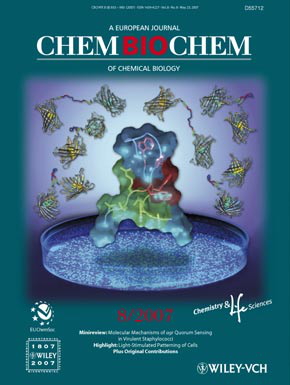
S.C. Alford, A.S. Abdelfattah, Y. Ding, and R.E. Campbell*, “A Fluorogenic Red Fluorescent Protein Heterodimer”. Chem. Biol., 2012, 19, 353-360. [Cover Art; Highlighted in Nature Methods; Supplementary Material; Funded by CIHR NHG 94487 and 99085, NSERC Discovery, NSERC CGSD3 to S.C.A., and Vanier CGS to A.S.A.]

40. FRET-based biosensors for multiparameter ratiometric imaging of Ca2+ dynamics and caspase-3 activity in single cells
Y. Ding, H-w. Ai, H. Hoi, R.E. Campbell*, “FRET-based biosensors for multiparameter ratiometric imaging of Ca2+ dynamics and caspase-3 activity in single cells”. Anal. Chem., 2011, 83, 9687–9693. [Supplementary Material; Funded by CIHR NHG 94487 and 99085 and NSERC Discovery]
39. A bacteria colony-based screen for optimal linker combinations in genetically encoded biosensors
A. Ibraheem, H. Yap, Y. Ding, R.E. Campbell*, “A bacteria colony-based screen for optimal linker combinations in genetically encoded biosensors”. BMC Biotechnol., 2011, 11, 105. [Open Access PDF; Supplementary Material; Funded by Alberta Cancer Board]
38. An Expanded Palette of Genetically Encoded Ca2+ Indicators
Y. Zhao, S. Araki, J. Wu, T. Teramoto, Y-F. Chang, M. Nakano, A.S. Abdelfattah, M. Fujiwara, T. Ishihara, T. Nagai, and R.E. Campbell*, “An Expanded Palette of Genetically Encoded Ca2+ Indicators”, Science, 2011, 333, 1888-1891. [Press release; Highlighted by C&EN Concentrates, Biophotonics, and Science Signaling; Supplementary Material; Funded by CIHR NHG 94487 and 99085, NSERC Discovery, Alberta Ingenuity Nanotechnology Scholarship to Y.Z., and Vanier CGS to A.S.A.]
37. A Monomeric Photoconvertible Fluorescent Protein for Imaging of Dynamic Protein Localization
H. Hoi, N.C. Shaner, M.W. Davidson, C.W. Cairo, J. Wang, R.E. Campbell*, “A Monomeric Photoconvertible Fluorescent Protein for Imaging of Dynamic Protein Localization”, J. Mol. Biol., 2010, 401, 776-791. [Supplementary Material; Funded by NSERC Discovery]
36. Circularly permuted monomeric red fluorescent proteins with new termini in the β-sheet
H.J. Carlson, D. Cotton, and R.E. Campbell*, “Circularly permuted monomeric red fluorescent proteins with new termini in the β-sheet”, Protein Sci., 2010, 19, 1490-1499. [Funded by NSERC Discovery, NSERC USRA to D.C., NSERC PGSM to H.J.C., and Alberta Ingenuity Scholarship to H.J.C.]
35. Fluorescent reporter proteins
R.E. Campbell and M.W. Davidson*, “Fluorescent reporter proteins”, Molecular Imaging with Reporter Genes. Eds. Sanjiv S. Gambhir and Shahriar S. Yaghoubi. Cambridge University Press, New York, NY, July 2010: 3 – 40. [Google book preview]
34. Designs and applications of fluorescent protein-based biosensors
A. Ibraheem and R.E. Campbell*, “Designs and applications of fluorescent protein-based biosensors”, Curr. Opin. Chem. Biol., 2010, 14, 30-36. [Funded by Alberta Cancer Board]
33. Molecular Imaging: Editorial Overview
R.E. Campbell* and C.J. Chang*, “Molecular Imaging: Editorial Overview“, Curr. Opin. Chem. Biol., 2010, 14, 1-2. [Co-editor for this Special issue of the journal which had 15 invited reviews.]
32. Engineered fluorescent proteins: innovations and applications
M.W. Davidson and R.E. Campbell*, “Engineered fluorescent proteins: innovations and applications”, Nat. Methods, 2009, 6, 713-717. [Invited Commentary for 5th Anniversary issue.]
31. Genetically encoded biosensors based on engineered fluorescent proteins
W.B. Frommer*, M.W. Davidson, R.E. Campbell* “Genetically encoded biosensors based on engineered fluorescent proteins”, Chem. Soc. Rev., 2009, 38, 2833-2841. [Supplementary Material]
30. Red fluorescent protein pH biosensor to detect concentrative nucleoside transport
D.E. Johnson, H-w. Ai, P. Wong, J.D. Young, R.E. Campbell*, and J.R. Casey* “Red fluorescent protein pH biosensor to detect concentrative nucleoside transport”, J. Biol. Chem., 2009, 284, 20499-20511. [Supplementary Material]
29. Fluorescent Protein-Based Biosensors: Modulation of Energy Transfer as a Design Principle
R.E. Campbell*, “Fluorescent Protein-Based Biosensors: Modulation of Energy Transfer as a Design Principle”, Anal. Chem., 2009, 81(15), 5972–5979. [Cover Art; Podcast]
28. An engineered tryptophan zipper-type peptide as a molecular recognition scaffold
Z. Cheng and R.E. Campbell*, “An engineered tryptophan zipper-type peptide as a molecular recognition scaffold”, J. Pept. Sci., 2009, 15, 523-532. [Supplementary Material]
27. Genetically encoded FRET-based biosensors for multiparameter fluorescence imaging
H.J. Carlson, R.E. Campbell*, “Genetically encoded FRET-based biosensors for multiparameter fluorescence imaging”, Curr. Opin. Biotechnol., 2009, 20, 19-27.
26. Fluorescent proteins
R.E. Campbell*, “Fluorescent proteins”, Scholarpedia J., 2008, 3(7), 5410. [Open access; Article accessed more than 95,000 times as of July 2014]
25. Fluorescent protein FRET pairs for ratiometric imaging of dual biosensors
H-w. Ai, K.L. Hazelwood, M.W. Davidson, and R.E. Campbell*, “Fluorescent protein FRET pairs for ratiometric imaging of dual biosensors”, Nat. Methods, 2008, 5, 401-403. [Supplementary Material; Highlighted in October 2008 issue of Biophotonics]
24. Hue-shifted monomeric variants of Clavularia cyan fluorescent protein: identification of the molecular determinants of color and applications in fluorescence imaging
H-w. Ai, S.G. Olenych, P. Wong, M.W. Davidson, and R.E. Campbell*, “Hue-shifted monomeric variants of Clavularia cyan fluorescent protein: identification of the molecular determinants of color and applications in fluorescence imaging”, BMC Biol., 2008, 6, 13. [Open Access PDF; Designated as a Highly Accessed article]
23. Teal fluorescent proteins: Characterization of a reversibly photoconvertible variant
H-w. Ai, and R. E. Campbell*, “Teal fluorescent proteins: Characterization of a reversibly photoconvertible variant”, Proc. SPIE, 2008, 6868, 68680D.
22. Computational prediction of absorbance maxima for a structurally diverse series of engineered green fluorescent protein chromophores
Q.K. Timerghazin, H.J. Carlson, C. Liang, R.E. Campbell,* and A. Brown*, “Computational prediction of absorbance maxima for a structurally diverse series of engineered green fluorescent protein chromophores”, J. Phys. Chem B, 2008, 112, 2533-2541. [Supplementary Material]
21. Identification of sites within a monomeric red fluorescent protein that tolerate peptide insertion and testing of corresponding circular permutations
Y. Li, A.M. Sierra, H.-w. Ai, and R.E. Campbell*, “Identification of sites within a monomeric red fluorescent protein that tolerate peptide insertion and testing of corresponding circular permutations”, Photochem. Photobiol., 2008, 84, 111–119. [Supplementary Material]
20. More than just pretty colors: the growing impact of fluorescent proteins in the life science
H-w. Ai and R.E. Campbell*, “More than just pretty colors: the growing impact of fluorescent proteins in the life sciences”, Biotechnology Focus (Bioscienceworld), 2007, issue 11, 16-18.
19. In vivo screening identifies a highly folded beta-hairpin peptide with a structured extension
Z. Cheng, M. Miskolzie, and R.E. Campbell*, “In vivo screening identifies a highly folded beta-hairpin peptide with a structured extension”, ChemBioChem, 2007, 8, 880-883. [Cover Art; Supplementary Material]

18. Exploration of new chromophore structures leads to the identification of improved blue fluorescent proteins
H.-w. Ai, N.C. Shaner, Z. Cheng, R.Y. Tsien, and R.E. Campbell*, “Exploration of new chromophore structures leads to the identification of improved blue fluorescent proteins”, Biochemistry, 2007,46, 5904 – 5910. [Featured on the cover of the June 2007 issue of Biophotonics]
17. Structural basis for reversible photobleaching of a green fluorescent protein homologue
J.N. Henderson, H.-w. Ai, R.E. Campbell, and S.J. Remington*, “Structural basis for reversible photobleaching of a green fluorescent protein homologue”, Proc. Natl. Acad. Sci. U.S.A., 2007, 14, 6672-6677. [Supplementary Material; Featured in June 2007 issue of Biophotonics and April 2007 Science Daily online]
16. Fluorescence-based characterization of genetically encoded peptides that fold in live cells: progress towards a generic hairpin scaffold
Z. Cheng and R.E. Campbell*, “Fluorescence-based characterization of genetically encoded peptides that fold in live cells: progress towards a generic hairpin scaffold”, Proc. SPIE, 2007, 6449, 64490S.
15. Directed evolution of a monomeric, bright, and photostable version of Clavularia cyan fluorescent protein: structural characterization and applications in fluorescence imaging
H-w. Ai, J.N. Henderson, S.J. Remington, and R.E. Campbell*, “Directed evolution of a monomeric, bright, and photostable version of Clavularia cyan fluorescent protein: structural characterization and applications in fluorescence imaging”, Biochem. J., 2006, 400, 531-540.
14. Assessing the Structural Stability of Designed β-Hairpin Peptides in the Cytoplasm of Live Cells
Z. Cheng and R.E. Campbell*, “Assessing the Structural Stability of Designed β-Hairpin Peptides in the Cytoplasm of Live Cells”, ChemBioChem, 2006, 7, 1147-1150.
13. Realization of β-lactamase as a versatile fluorogenic reporter
R.E. Campbell*, “Realization of β-lactamase as a versatile fluorogenic reporter”, Trends Biotech., 2004, 22, 208-211.
Postdoctoral research at the University of California, San Diego:
12. Autofluorescent Proteins with Excitation in the Optical Window for Intravital Imaging in Mammals
M.Z. Lin, M.R. McKeown, H.-L. Ng, T.A. Aguilera, N.C. Shaner, R.E. Campbell, S.R. Adams, L.A. Gross, W. Ma, T. Alber, R.Y. Tsien*, “Autofluorescent Proteins with Excitation in the Optical Window for Intravital Imaging in Mammals”, Chem. Biol., 2009, 16, 1169-1179.
11. Improved monomeric red, orange, and yellow fluorescent proteins derived from Discosoma red fluorescent protein
N.C. Shaner, R.E. Campbell, P.A. Steinbach, B.N.G. Giepmans, A.E. Palmer, and R.Y. Tsien*, “Improved monomeric red, orange, and yellow fluorescent proteins derived from Discosoma red fluorescent protein”, Nat. Biotechnol., 2004, 22, 1567-1572.
10. Creating New Fluorescent Probes for Cell Biology
J. Zhang, R.E. Campbell, A.Y. Ting and R.Y. Tsien*, “Creating New Fluorescent Probes for Cell Biology”, Nat. Rev. Mol. Cell Biol., 2002, 3, 906-918.
9. A Monomeric Red Fluorescent Protein
R.E. Campbell, O. Tour, A.E. Palmer, P.A. Steinbach, G.S. Baird, D.A. Zacharias and R.Y. Tsien*, “A Monomeric Red Fluorescent Protein”, Proc. Natl. Acad. Sci. U.S.A., 2002, 99, 7877-7882.
8. New Biarsenical Ligands and Tetracysteine Motifs for Protein Labeling in Vitro and in Vivo: Synthesis and Biological Applications
S.R. Adams, R.E. Campbell, L.A. Gross, B.R. Martin, G.K. Walkup, Y. Yao, J. Llopis and R.Y. Tsien*, “New Biarsenical Ligands and Tetracysteine Motifs for Protein Labeling in Vitro and in Vivo: Synthesis and Biological Applications”, J. Am. Chem. Soc., 2002, 124, 6063-6076.
7. Reducing the Environmental Sensitivity of Yellow Fluorescent Protein: Mechanism and Applications
O. Griesbeck, G.S. Baird, R.E. Campbell, D.A. Zacharias and R.Y. Tsien*, “Reducing the Environmental Sensitivity of Yellow Fluorescent Protein: Mechanism and Applications”, J. Biol. Chem., 2001, 276, 29188-29194.
Graduate research at the University of British Columbia:
6. The Structure of UDP-N-Acetylglucosamine 2-Epimerase Reveals Homology to Phosphoglycosyl Transferases
R.E. Campbell, S.C. Mosimann, M.E. Tanner*, and N.C.J. Strynadka*, “The Structure of UDP-N-Acetylglucosamine 2-Epimerase Reveals Homology to Phosphoglycosyl Transferases”,Biochemistry, 2000, 39, 14993-15001.
5. The First Structure of UDP-Glucose Dehydrogenase Reveals the Catalytic Residues Necessary for the Two-fold Oxidation
R.E. Campbell, S.C. Mosimann, I. van de Rijn, M. E. Tanner, and N.C.J. Strynadka*, “The First Structure of UDP-Glucose Dehydrogenase Reveals the Catalytic Residues Necessary for the Two-fold Oxidation”, Biochemistry, 2000, 39, 7012-7023.
4. UDP-Glucose Analogues as Inhibitors and Mechanistic Probes of UDP-Glucose Dehydrogenase
R.E. Campbell and M.E. Tanner*, “UDP-Glucose Analogues as Inhibitors and Mechanistic Probes of UDP-Glucose Dehydrogenase”, J. Org. Chem., 1999, 64, 9487-9492.
3. Covalent Adduct Formation with a Mutated Enzyme: Evidence for a Thioester Intermediate in the Reaction Catalyzed by UDP-Glucose Dehydrogenase
X. Ge, R.E. Campbell, I. van de Rijn, and M.E. Tanner*, “Covalent Adduct Formation with a Mutated Enzyme: Evidence for a Thioester Intermediate in the Reaction Catalyzed by UDP-Glucose Dehydrogenase”, J. Am. Chem. Soc., 1998, 120, 6613-6614.
2. Uridine diphospho-alpha-D-gluco-hexodialdose: Synthesis and kinetic competence in the reaction catalyzed by UDP-glucose dehydrogenase
R.E. Campbell and M.E. Tanner*, “Uridine diphospho-alpha-D-gluco-hexodialdose: Synthesis and kinetic competence in the reaction catalyzed by UDP-glucose dehydrogenase”, Angew. Chem. Int. Ed. Eng. 1997, 36, 1520-1522.
1. Properties and kinetic analysis of UDP-glucose dehydrogenase from group A streptococci. Irreversible inhibition by UDP-chloroacetol
R.E. Campbell, R.F. Sala, I. van de Rijn and M.E. Tanner*, “Properties and kinetic analysis of UDP-glucose dehydrogenase from group A streptococci. Irreversible inhibition by UDP-chloroacetol”, J. Biol. Chem., 1997, 272, 3416-22.